About Rouse EyeCare Center
Serving Lacey and the surrounding communities, we offer comprehensive eye health services for all members of your family. We know how much your eye health and appearance mean to the quality of your life. Dr. Craig Rouse and our staff are committed to excellence and serving your complete eye care needs.
Visit us at Rouse EyeCare Center to find the latest in fashion eyewear and lens technology. Our experienced staff looks forward to helping you find the best pair of eyewear or contact lenses to meet your visual needs.
If you are new to our website, we invite you to explore and discover all there is to learn about your vision. We also encourage you to subscribe to our newsletter. In addition, become a member of our website! Members enjoy exclusive access to special promotions, articles, resources, and eye health facts specifically chosen by Craig Rouse.
We look forward to welcoming you to our office at Rouse EyeCare Center soon!


Our Eye Care Services

Comprehensive Eye Exams
From updating your eyeglasses prescription to detection and treatment of eye diseases, comprehensive eye exams are important for continued visual health.

Contact Lens Exams
During a contact lens exam, your eye doctor will check if you are a good candidate for contacts and find the best type of contacts for your needs..

Dry Eyes
Do you have dry, itchy, gritty-feeling eyes? Dry eye syndrome is a very common condition. We offer dry eye treatments at our eye care clinic.
Clear vision is an essential part of a child’s healthy development and learning. Regular pediatric eye exams can help detect and treat common issues.
Children with myopia are at higher risk for potentially sight-threatening vision problems. Save your child’s vision with myopia management.
Eye diseases such as glaucoma and macular degeneration can cause severe loss of vision if not diagnosed and treated early. Our eye care team can help.
If cataracts go untreated, they can cause total blindness in the affected eye. We can help with co-management of your cataract removal surgery.
Macular degeneration can cause severe central vision loss if not detected and treated early on. We can help preserve your vision.
Astigmatism can cause vision problems such as blurred or double vision at all distances. Find out what you can do to correct astigmatism and see your best.
The ups and downs of diabetes can be difficult to navigate. How can diabetes affect your eyes? How can you and your eye doctor keep them healthy?
While vision can change drastically in the senior years, these changes shouldn't have to impair one's quality of life. The earlier these problems are detected and treated, the more likely proper eye care can help you retain good vision.
Eyeglasses & Frames

Designer Frames
Our extensive optical section offers a wide variety of eyeglass frames in every style, material & design. Come visit us today to see for yourself!
Our expert optical team can find just the right pair of glasses for you to be confident and look your best.
Lens coatings improve visual comfort, make it easier to clean your glasses and ensure your lenses last longer. Coatings include anti-scratch, anti-reflective, photochromatic and UV / blue light filters.
Contact Lenses

Contact Lens Fitting
We offer a wide range of contact lens options from dailies, monthly to multifocal contact lenses for crystal clear vision and superior comfort.
During a contact lens exam, your eye doctor will check if you are a good candidate for contacts and find the best type of contacts for your needs.
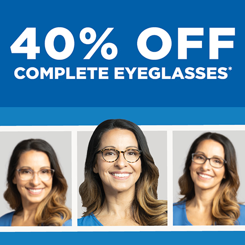
40% Off Complete Eyeglasses
*Requires purchase of a complete prescription pair, including frame and lenses. Does not include sunglass frames, Barton Perreira, Cartier, Cazal, Chanel, Cutler and Gross, Dior, Dita Lancier, Fendi, Gucci, ic!Berlin, l.a. Eyeworks, Maui Jim, Mykita, Nifties, Oakley, Oliver Peoples, Persol, Ray-Ban, Robert Marc, Salt, Salvatore Ferragamo, Skaga, Silhouette, Tom Ford, WOOW, accessories, contact lenses, or medical procedures. Cannot be combined with any other discounts, promotions, or insurance plans. Not valid on previous orders. Other restrictions may apply. See practice for full details. Offer valid 04/08/2024-06/16/2024. 24AEG-729313